
Medically reviewed by Dr. Neha Yadav — Infertility Specialist & IVF Consultant at Renew Healthcare, Salt Lake, Kolkata
Last updated: September 2026
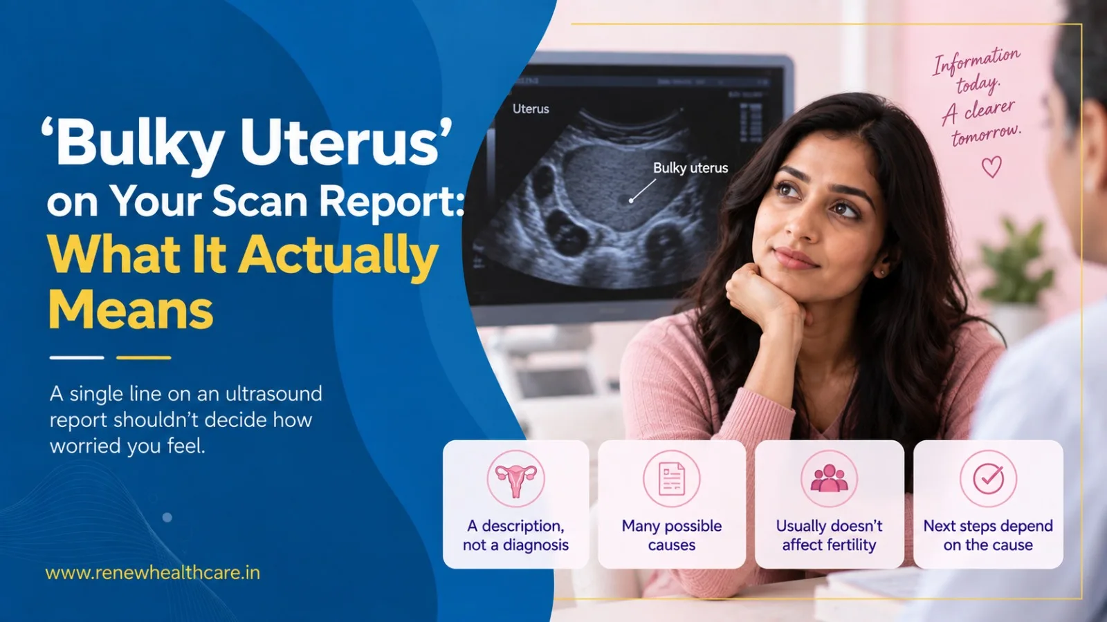
In brief: A single line on an ultrasound report should not decide how worried you feel. Bulky uterus is a descriptive term meaning the uterus measured larger than the reference range that scan centre uses — not a diagnosis. Whether it affects conception depends almost entirely on the cause behind the size, and most women with a mildly bulky uterus and no other findings conceive without any uterus-specific treatment.
You went in for a routine scan — maybe for irregular periods, maybe as part of a fertility work-up — and the report came back with one unexpected phrase: bulky uterus. No one explained it at the desk, and a quick search online turns up a mix of reassurance and alarm, sometimes on the same page. So what does this term actually mean, and should it change your fertility plans?
Key takeaways
- Bulky uterus is a descriptive ultrasound term, not a diagnosis — it simply means the uterus measured larger than the typical reference range used by that scan centre.
- There is no single, universally agreed cutoff for a normal uterus size — reference ranges vary by study population, parity and even the ultrasound technique used.
- The most common causes — fibroids, adenomyosis, prior pregnancy, and the normal hormonal swing of the menstrual cycle — differ enormously in how much they matter for fertility.
- Whether a bulky uterus affects conception depends almost entirely on its underlying cause, not on the label itself.
- Most women with a mildly bulky uterus and no other findings go on to conceive without any uterus-specific treatment.
What bulky uterus actually means
A uterus is called bulky when it measures larger than the reference range the sonographer or radiologist is using. It is a description of size, not a diagnosis — comparable to a doctor noting that your blood pressure reading was higher than average before working out why.
Here is the honest part that competitor content often skips: there is no single global cutoff for what counts as normal. Reference ranges differ by study population, by whether a woman has been pregnant before, and even by which day of her cycle the scan was done. A recent multicentre European study confirmed this directly, finding that no universally accepted ultrasound definition of a normal uterus exists in the literature. That is not a gap in your understanding; it is a genuine gap in medical standardisation.
What usually causes it
1. A uterus that has carried a pregnancy before
Childbirth permanently and normally increases uterine size. This is not a condition to treat — it is simply how the organ changed.
2. Timing within the menstrual cycle
The uterine lining thickens and thins across the month. A scan done later in the cycle can look bulkier than one done earlier, with nothing pathological happening at all.
3. Fibroids
These non-cancerous growths in the uterine wall are extremely common and, in most cases, do not affect fertility. What matters is location: fibroids that push into the uterine cavity are far more relevant than small ones sitting on the outer wall.
4. Adenomyosis
This is where uterine-lining tissue grows into the muscular wall. Its link to fertility and IVF outcomes is still being actively researched — some studies show an association with lower success rates, others show none. We flag this honestly rather than presenting it as settled science.
| Underlying cause | How common | Typical fertility relevance |
|---|---|---|
| Prior pregnancy (parity) | Very common | None — a normal, permanent physiological change, not a fertility issue at all. |
| Adenomyosis | Common in women 30 and over | Evidence is still evolving; some studies link diffuse adenomyosis to lower IVF success, others show no difference. |
| Fibroids (myomas) | Very common (up to roughly 70–80% of women by menopause, per pooled estimates) | Depends heavily on location. Submucosal fibroids distort the cavity and matter most; small subserosal fibroids usually do not. |
| Hormonal or cycle-phase timing | Common, especially mid-cycle scans | None — the uterus normally thickens across the cycle; a scan on a different day may look different. |
| Endometrial hyperplasia | Uncommon in reproductive years | Needs prompt evaluation; not typical in a young woman with regular cycles. |
Does it actually affect your fertility?
Usually, the label itself changes nothing. What changes your fertility odds is the specific cause behind the size — particularly whether it distorts the cavity where an embryo would implant. A mildly bulky uterus with a normal cavity, no heavy bleeding and no pain is, in most cases, an incidental finding rather than a fertility diagnosis.
Myth vs. fact
| Myth | Fact |
|---|---|
| A bulky uterus means I have a tumour or cancer. | In reproductive-age women with regular cycles, malignancy is a rare cause. Most bulky-uterus findings trace back to fibroids, adenomyosis or normal cycle-day variation. |
| A bulky uterus always causes infertility. | The label alone predicts very little. What matters is the specific cause, its size and location, and whether it distorts the uterine cavity. |
| If my report says bulky uterus, I need surgery immediately. | Most cases are managed with monitoring, symptom-directed treatment or targeted therapy — not surgery. Surgery is reserved for specific indications such as cavity-distorting fibroids. |
| Every clinic will measure my uterus the same way and get the same result. | Reference ranges differ across studies and scan centres, and measurements shift with parity, cycle day and technique — which is why the same uterus can be read differently on different days. |
What to do next
- Ask your doctor which reference range was used, and on which cycle day the scan was done.
- Request the specific cause — fibroid, adenomyosis, cycle timing or physiological — rather than accepting bulky as a final answer.
- If fibroids are involved, ask specifically about their location relative to the uterine cavity.
- Track any symptoms — heavy bleeding, pelvic pain, irregular cycles — to bring to your next appointment.
- Avoid rushing into surgery or treatment before the underlying cause is confirmed.
Frequently asked questions
Is a bulky uterus the same as fibroids?
No. Bulky uterus describes overall size; fibroids are one possible cause among several. A report can say bulky uterus with or without fibroids being present.
Can I still get pregnant naturally with a bulky uterus?
In most cases, yes. Unless the underlying cause is distorting the uterine cavity or significantly disrupting the lining, natural conception is usually still possible.
Does a bulky uterus affect IVF success?
It depends on the cause. A mildly bulky uterus with a normal cavity and no other findings is not generally treated as a red flag in IVF planning; adenomyosis and cavity-distorting fibroids warrant individual discussion with your specialist.
What tests should follow a bulky uterus finding?
Typically a detailed transvaginal ultrasound (sometimes with 3D or saline infusion views), a review of cycle timing, and correlation with symptoms such as heavy bleeding or pain. Further tests depend on what these show.
Should I be alarmed if I have no symptoms at all?
An incidental, symptom-free finding on a routine scan is common and, by itself, is rarely urgent. It is still worth discussing with your gynaecologist so the cause is understood rather than assumed.
A report is a starting point, not a verdict
If your scan mentions a bulky uterus, Dr. Neha Yadav can help you understand the specific cause behind it and whether it has any bearing on your fertility plans — with a clear, evidence-based explanation before any treatment is discussed. Consultations are available at the Renew Healthcare clinic in Salt Lake, Kolkata.
References and evidence notes
- Uterine size and shape reference ranges vary by study population and technique; pooled nulliparous estimates cluster around 7–8 cm length, 4–5 cm width and 3–4 cm thickness, with larger dimensions in parous women.
- Normal UteRus asSEssment (NORMS) study — multicentre European cohort establishing 3D ultrasound percentile ranges for the normal nulliparous uterus, noting that no universally accepted ultrasound definition of a normal uterus exists. Fertility and Sterility, 2025.
- Fibroid prevalence estimates of up to roughly 70–80% by age 50 are drawn from pooled and secondary sources rather than a single primary epidemiological study.
- ASRM Practice Committee guidance on the management of uterine fibroids in relation to fertility — location-dependent impact of submucosal versus subserosal fibroids on implantation.
- ESHRE guidance and current literature on adenomyosis and reproductive outcomes — evidence on IVF impact remains mixed and is flagged here as an evolving area rather than settled consensus.
- Uterine factors account for an estimated 3–5% of infertility cases, per secondary-source citation.